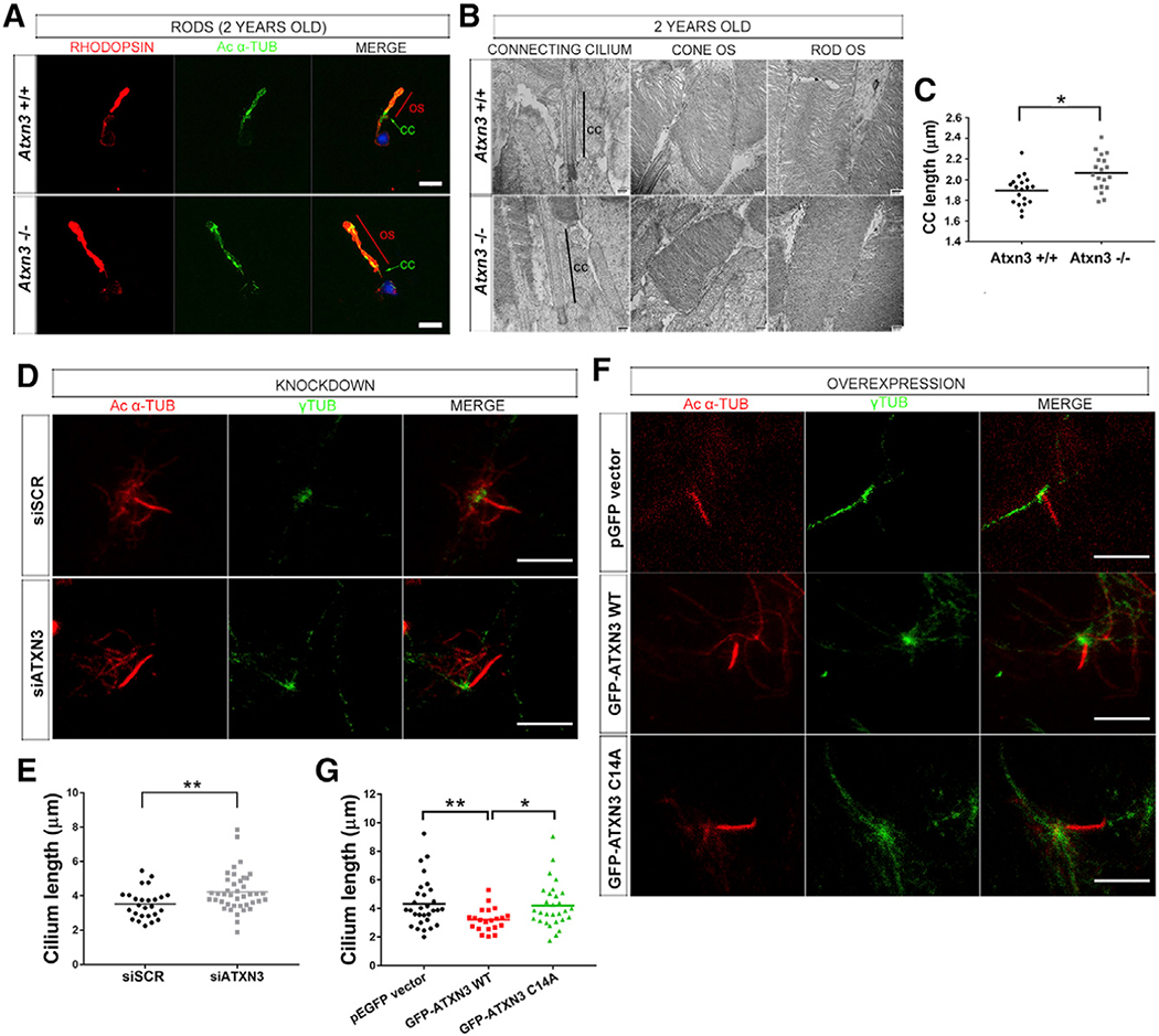
Figure 3. ATXN3 Regulates the Length of the Cilium in PhR Cells.
(A–C) Elongation of the OS (contains the axoneme) and CC (connecting cilium) in Atxn3 KO PhRs, as shown in (A), by rhodopsin (red) and acetylated-α-tubulin (green) detection in isolated rods from 2-year-old WT (Atxn3+/+) and KO (Atxn3−/−) mouse retinas; nuclei were labeled with DAPI (blue) (scale bar, 10 μm); and in (B), by transmission electron microscopy (TEM), where the connecting cilium is indicated by a black line. No other apparent morphological differences were detected (representative images). (C) Mean CC length was 1.897 μm for WT and 2.066 μm for KO (19 TEM pictures, 3 animals per genotype). Two-way ANOVA test (*p < 0.05).
(D–G) The length of primary cilia in ARPE-19 cells is modulated by ATXN3 levels. (D and E) Depletion of endogenous ATXN3 by siATXN3 transfection in human ARPE-19 cells results in longer primary cilium (mean 4.222 μm in siATXN3 cells, n = 41) than that in controls (mean 3.523 μm in siSCR cells, n = 26). Mann-Whitney test (**p < 0.01). (F and G) Starved ARPE-19 cells overexpressing GFP-ATXN3 WT Q22 produce shorter cilia (mean, 3.211 μm) than cells with GFP-ATXN3 C14A mutant (4.208 μm) or the pEGFP empty vector (mean 4.318 μm). Only GFP-positive cells (n > 20–30 per condition) were analyzed (see Figure S5B). Mann-Whitney test (*p < 0.05, **p < 0.01). In (D) and (F), Ciliary microtubules were detected with acetylated α-tubulin (red) and basal bodies with γ-tubulin (green). Scale bar, 5 μm.

