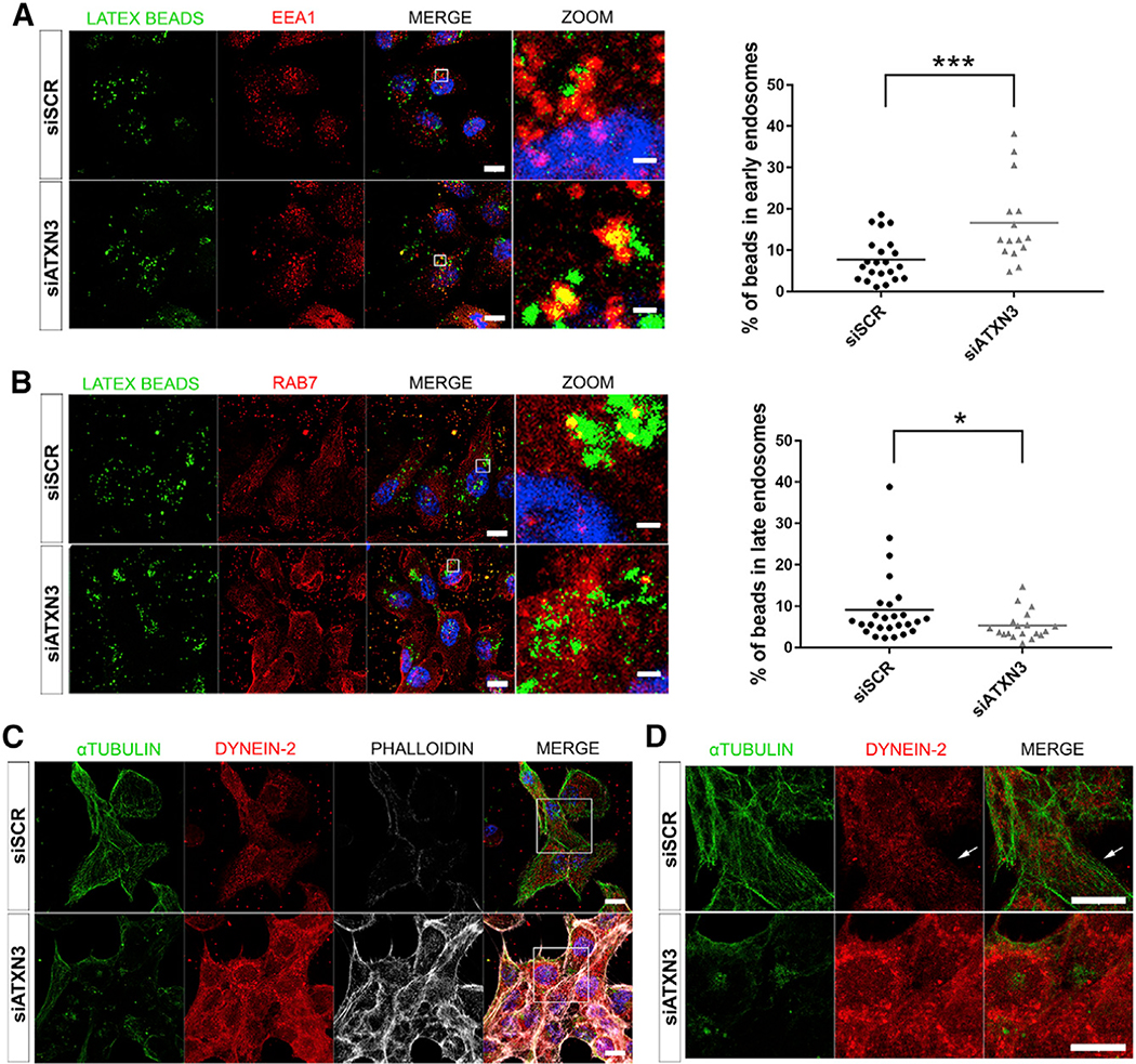
Figure 6. Depletion of ATXN3 in ARPE-19 Cells Causes an Increase of Early Endosomes, Decrease of Late Endosomes, and Alterations in the Cytoskeleton Organization.
(A and B) Immunofluorescent detection of early endosomes (EEA1, red) (A) and late endosomes (RAB7, red) (B) in ATXN3-depleted ARPE-19 cells after incubation with fluorescent latex beads (green) to induce phagocytosis. Quantification of the percentage of the beads within these vesicles shows a significant increase of early endosomes and a decrease of late endosomes. Mann-Whitney test (*p < 0.05, ***p < 0.001, n > 15 cells per group).
(C) Knockdown of ATXN3 in ARPE-19 phagocytic cells alters cytoskeleton organization, as shown by microtubule disorganization (α-tubulin, green), dynein-2 disarray (red), and F-actin increase (phalloidin, gray).
(D) Zoom-in at the central region of images in (C). White arrows point to dynein-2 dots trailing microtubule filaments in control cells (3D visualization in Videos S5 and S6). Nuclei were labeled with DAPI (blue). Scale bar in (A)–(C),10 μm.

