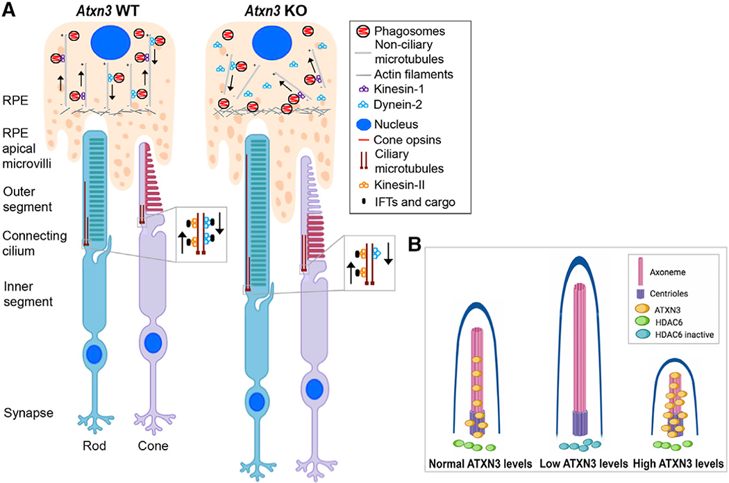
Figure 7. Model for the Molecular Role of ATXN3 in the PhRs and RPE Cells Based on the Phenotype Observed in Atxn3 KO Mouse Retinas.
(A) Rods and cones are PhR cells with a highly specialized sensory cilium, the OS, which contains ordered membranous stacks packed with proteins involved in phototransduction (e.g., rod and cone opsins), which are produced at the IS and must traffic through the ciliary gate. Sensory cilia have two centrioles at the base, from which the corresponding ciliary microtubules grow into the connecting cilium and the axoneme (well within the OS). The intraciliary transport (IFT) of cargo proteins is bidirectional and relies on microtubule-associated motors, kinesins, and dyneins, which are responsible for anterograde and retrograde transport. RPE cells produce microvilli that phagocyte the tips of the OS for renewal of photoreception and phototransduction machinery. Actin filaments, ezrin, and dynamin-2 are involved in RPE phagocytosis. Phagosomes/early endosomes are formed in the apical zone of the RPE, and their maturation process into late endosomes and fusion to the lysosomes at the basal RPE zone involve non-ciliary microtubule-dependent transport (retrograde direction).
(B) Our model proposes that in the retina, ATXN3 modulates the retrograde transport mediated by both ciliary and non-ciliary microtubules by regulation of HDAC6 activity. In the absence or low levels of ATXN3, the following events may occur: (1) the levels of KEAP1 are decreased, which, in turn, increase the levels of p62 and thus inhibits HDAC6; (2) as a result, the pool of acetylated tubulin increases and the polymerization of microtubules is enhanced; (3) the cilium and OS of PhRs increase in length and the retrograde transport is altered, thus resulting in cone opsin mislocalization, phagosome maturation delay, and microtubule disorganization. When overexpressed, ATXN3 localization into the basal body and axoneme increases, where it may enhance microtubule depolymerization and resorption, resulting in shorter cilia (images with biorender.com).

