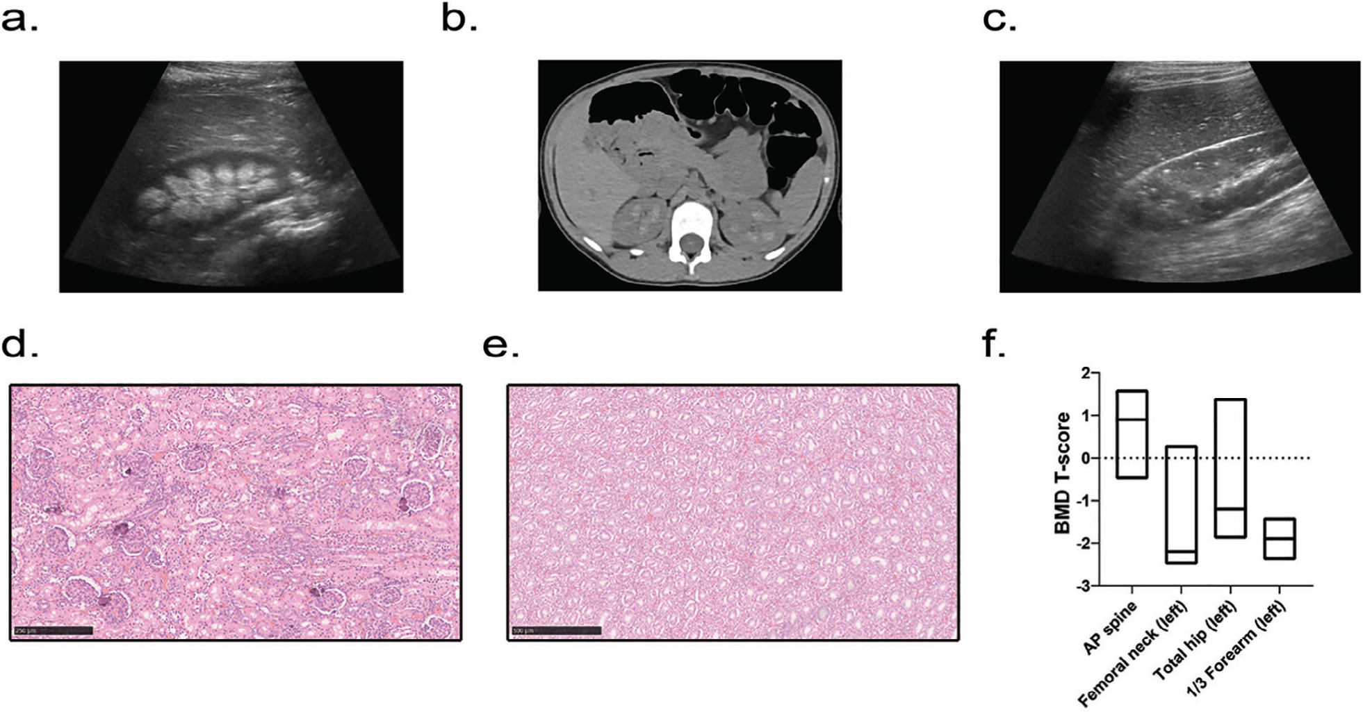
Fig 1.

Human phenotype. (A–C) Renal imaging. (A) Renal ultrasound showing medullary nephrocalcinosis (patient 15, right kidney, aged 9 years 8 months). (B) Computed tomography of the abdomen revealing bilateral calcification of the renal pyramids (patient 6, aged 8 years 3 months). (C) Renal ultrasound revealing punctate hyperechogenic foci within the renal cortex (patient 12, right kidney, aged 7 years 11 months). (D, E). Renal histology of patient 1’s deceased sibling revealing foci of calcification within the renal cortex (D), including glomeruli, with absent calcification within the medulla (E). (F) Areal bone mineral density T-scores in adults; data are displayed as box plots denoting median value and interquartile range.
