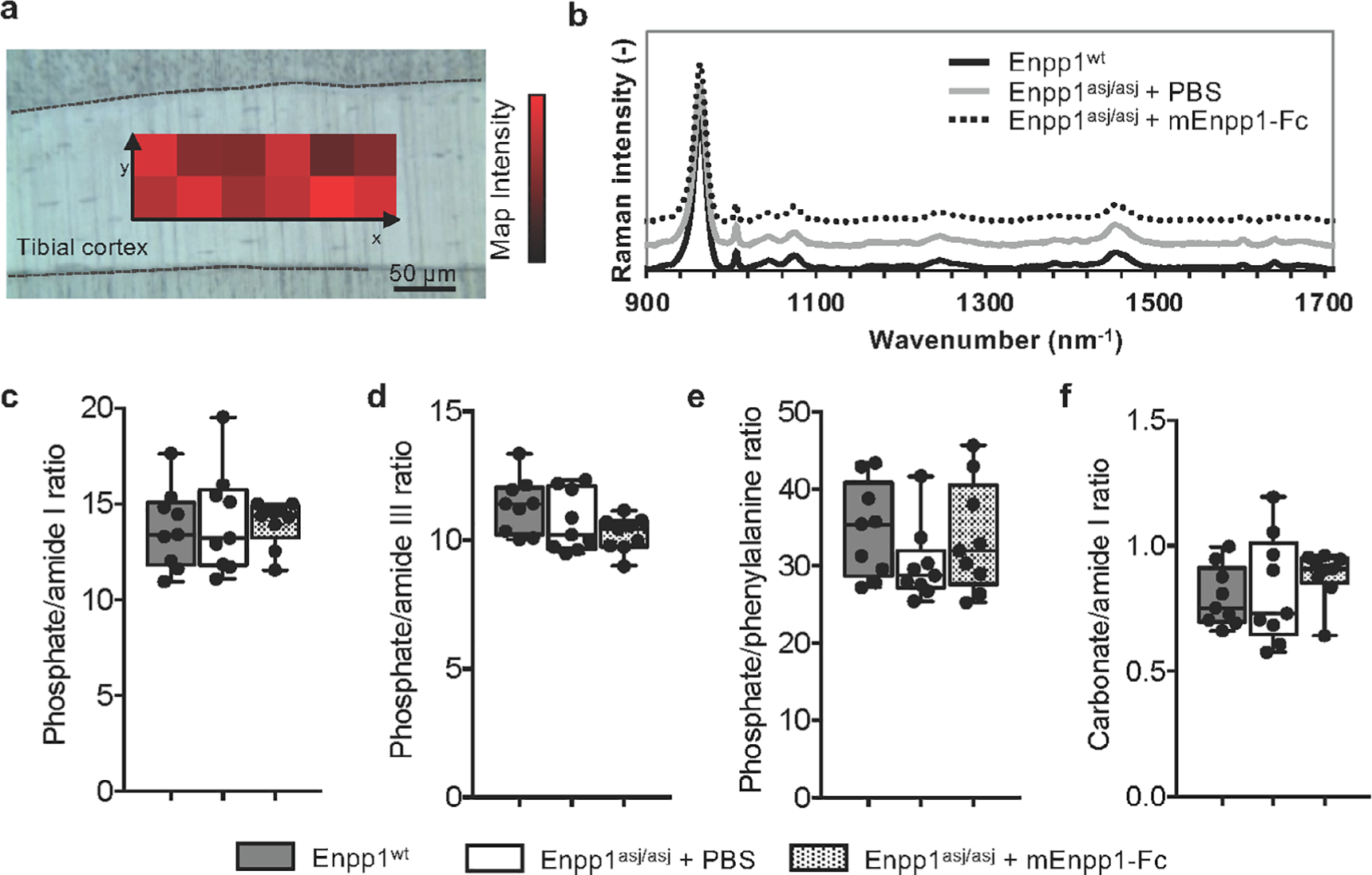
Fig 5.

Raman spectroscopy measurements of WT and Enpp1asj/asj mice treated with vehicle or mEnpp1-Fc. (A) Rectangular maps were acquired on the lateral and medial tibial cortex of each specimen. (B) Comparing the normalized average Raman spectra of each group indicates no differences in peak size or location. (C–F) Raman metrics based on the peak areas were assessed in male and female mice and did not show any statistical difference in (C) phosphate/amide I ratio (p = .67 between females, p = .85 between males), (D) phosphate/amide III (p = .22 between females, p = .53 between males), (E) phosphate/phenylalanine (p = .11 between females, p = .91 between males), and (F) carbonate/amide I (p = .76 between females, p = .18 between males), suggesting that neither the mutation nor the therapy significantly affected the bone matrix composition at the tissue level.
