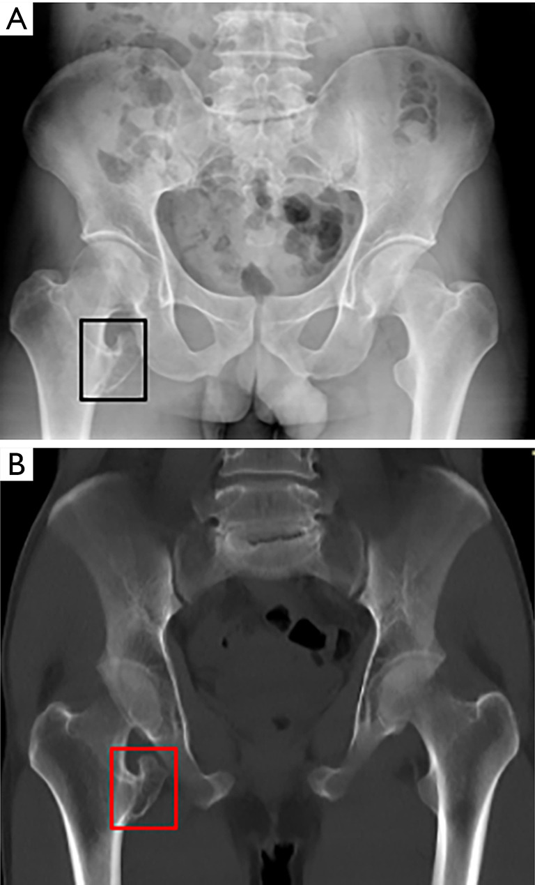
Figure 1.

X-ray and CT MIP reconstruction results. (A,B) Pelvic X-ray (A) and coronal CT MIP reconstruction (B) (slice thickness: 10 mm) show the presence of an osteochondroma of the lesser trochanter and narrowing of the distance between the lesser trochanter and the ischial tuberosity (black and red frames). CT, computed tomography; MIP, maximum intensity projection.
