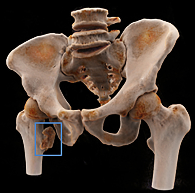
Figure 2.

CR image from the CT data at the coronal plane. This CR image contained detailed anatomical information of the osteochondroma in the right lesser trochanter bone and the relationship between the osteochondroma and the parent bone (blue frame). CR, cinematic rendering; CT, computed tomography.
