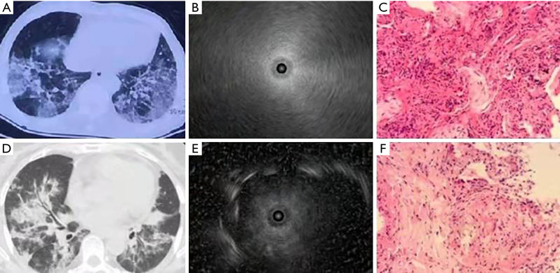
Figure 3.
The EBUS images of alveolar lesions on chest CT imaging. (A) Ground-glass lesions with bilateral distribution on chest CT imaging; (B) a blizzard sign; (C) organizing pneumonia in pathology (HE, ×100); (D) consolidation at the middle lobe of the right lung with an air bronchogram on chest CT imaging; (E) low-echo areas with or without a clear boundary within the solid shadow on ultrasound using an ultrasound probe; (F) granulomatous inflammation was considered to sarcoidosis in pathology (HE, ×100). EBUS, endobronchial ultrasound; CT, computed tomography; HE, hematoxylin and eosin staining.

