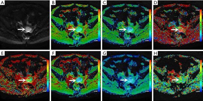
Figure 2.
Amide proton transfer-weighted imaging (APTWI) and multimodel diffusion-weighted imaging (DWI) images of an endometrioid adenocarcinoma (EA) patient (53-year-old woman, grade 3, FIGO II, and Ki-67=60%) (arrows indicate lesions). (A) Map of DWI (b=1,000 s/mm2), (B) pseudo–colored map of apparent diffusion coefficient (ADC), (C) pseudo–colored map of diffusion coefficient (D), (D) pseudo-colored map of pseudo-diffusion coefficient (D*), (E) pseudo–colored map of perfusion fraction (f), (F) pseudo–colored map of distributed diffusion coefficient (DDC), (G) pseudo–colored map of water molecular diffusion heterogeneity index (α), and (H) pseudo–colored map of magnetization transfer ratio asymmetry [MTRasym (3.5 ppm)].

