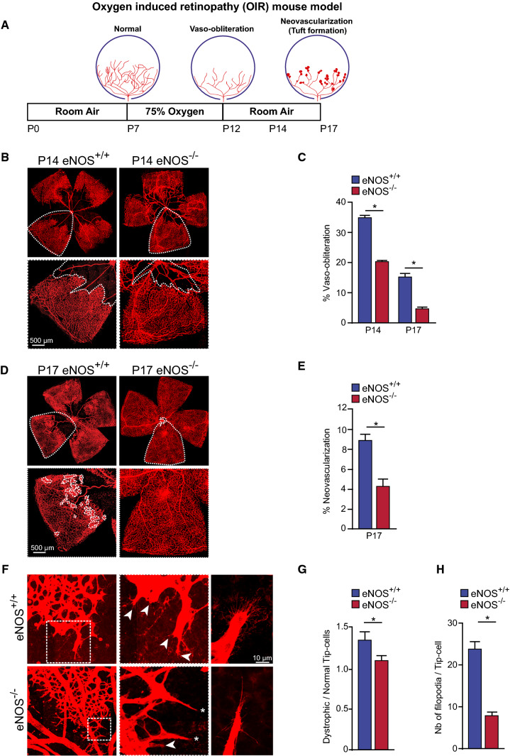
Fig. 6.
eNOS is a modulator of pathological neovascularization in oxygen-induced retinopathy. A Schematic representation of oxygen-induced retinopathy (OIR) mouse model. From postnatal day 7 (P7) to P12, mice are exposed to hyperoxic conditions (75% O2), leading to vaso-obliteration. Mice are then returned to room air resulting in hypoxia-driven revascularization and pathologic neovascularization, and tuft formation. B Representative high-resolution imaging of Isolectin B4–stained mouse retina at P14. C Quantification of vaso-obliteration (VO) is expressed as the central avascular areas divided by the total area of the retina for each strain. D Representative high-resolution imaging of Isolectin B4–stained mouse retina at P17. Areas of neovascularization are outlined in lower panels. E Quantification of neovascularization area in mouse strains. F Representative images of normal (asterisks) and dystrophic cells (arrowheads) at the vascular front of the retina in eNOS+/+ and eNOS−/− mice. G Quantification of the ratio of dystrophic tip-cells to normal tip-cells. H Measurement of the number of filopodia per tip-cell. Results are displayed as mean values ± SEM. *p < 0.05 (n = 6 for P14 and n = 10 for P17 eNOS+/+ mice; n = 22 for P14 and n = 34 for P17 eNOS−/− mice)

