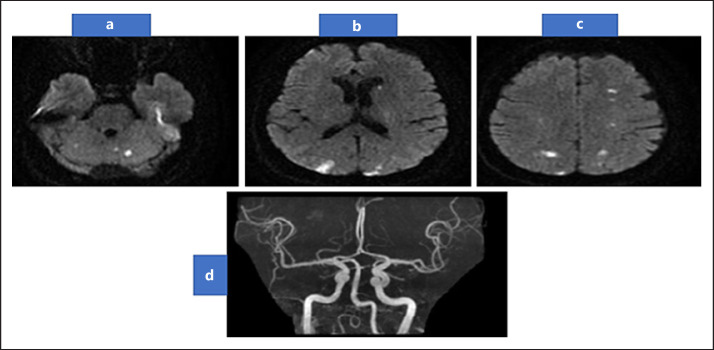
Fig. 1.
DWI-MRI brain showing multiple small foci of restricted diffusion on cortical, subcortical regions of frontal, parietal, and occipital hemispheres (a–b). In addition to both cerebellar hemisphere (c), with normal MRA (d). DWI, diffusion-weighted imaging; MRI, magnetic resonance imaging; MRA, magnetic resonance angiography.

