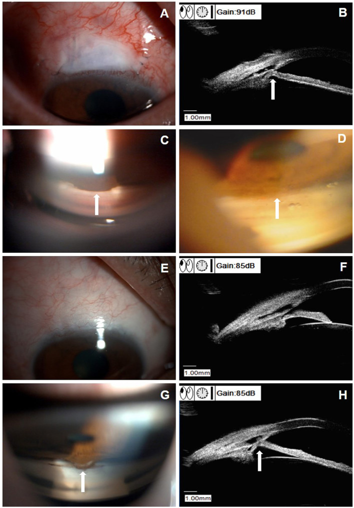
Figure 3.
Clinical presentations of a patient (patient six in Supplemental Table 1) with peripheral anterior synechiae (PAS) after CLASS. (A) Slit-lamp examination (DC-4, Topcon Corporation, magnification 16X) of a 61-year-old male patient at 3 weeks after CLASS. (B) Ultrasound biomicroscopy (UBM) examination indicated PAS (white arrow). (C) Gonioscopy examination confirmed PAS around the treating area (white arrow). (D) After Nd:YAG laser synechiolysis treatment, the iris retreated from the trabeculodescemetic window (TDW) (white arrow). (E) Slit-lamp examination of the patient at 6 months after CLASS showed no obvious filtering bleb. (F) UBM examination at 6 months after CLASS showed no PAS. (G) Gonioscopy examination at 12 months after CLASS showed a second PAS (white arrow). (H) UBM examination confirmed a second PAS (white arrow) at 12 months after CLASS.

