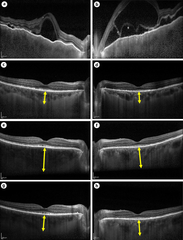
Fig. 2.
EDI-OCT images during patient follow-up. Left column shows EDI-OCT images of the right eye, and the right column shows those of the left eye. a, b Before treatment, SRD, choroidal folds, and marked choroidal thickening are observed (CT cannot be measured). c, d After the second steroid pulse treatment, serous retinal detachment disappeared and CT decreased (CT: right eye 390 μm, left eye 330 μm). e, f Nivolumab administration was resumed, and there was no significant change in the first administration, but CT increased and inflammation recurred in the second administration (CT: right eye 665 μm, left eye 485 μm). g, h One week after STTA was performed on both eyes for relapse. Choroidal thickening in both eyes improved. (CT: right eye 370 μm, left eye 350 μm). EDI-OCT, enhanced-depth imaging optical coherence tomographic; CT, choroidal thickness; STTA, subtenon injection of triamcinolone acetonide.

