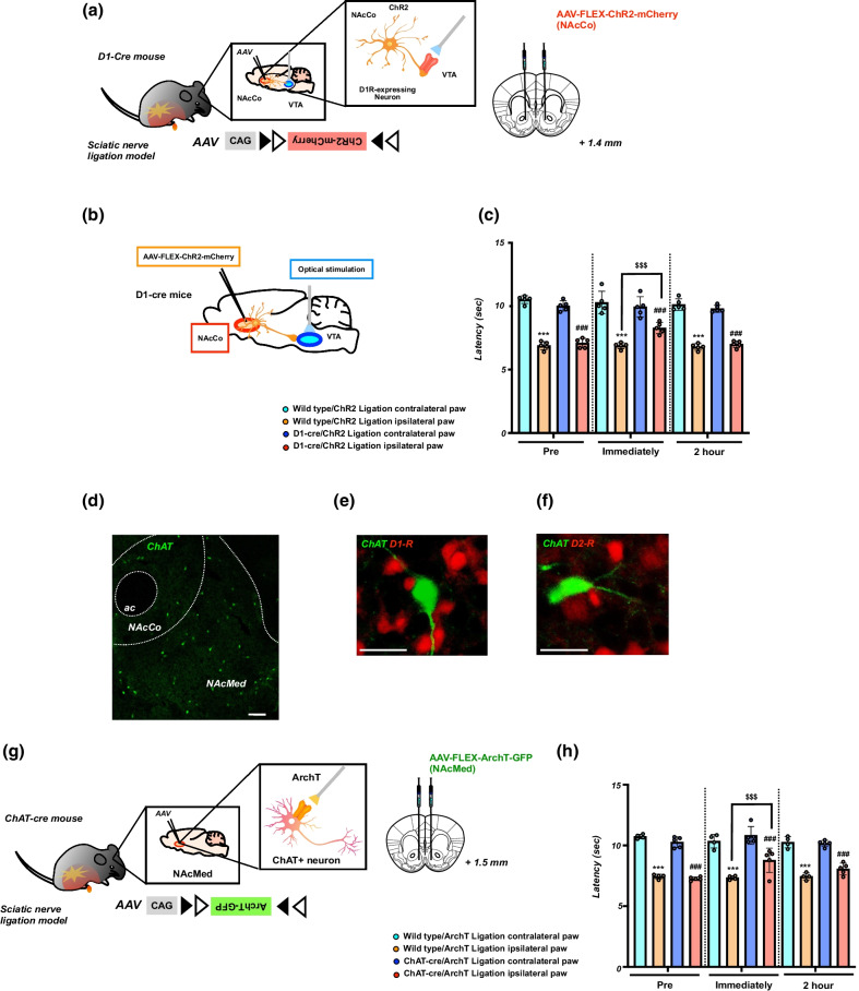
Fig. 5.
Effect of optical stimulation of D1-receptor-expressing neurons in the NAc projecting to the VTA and optical inhibition of cholinergic neurons in the NAc on neuropathic pain. a Schematic showing that AAV-Flex-ChR2-mCherry was microinjected into the NAcCo of D1-cre mice. b Schematic diagram showing the experimental design. c Effects of optical stimulation of D1-receptor-expressing neurons projecting to the VTA. Latency in the response to thermal stimulation under a neuropathic pain-like state by optical stimulation of the VTA in Wild-type/ChR2 and D1-cre/ChR2 mice. Data are presented as the mean ± SEM of 5 animals. ***p < 0.001 vs. Wild-type/ChR2 ligation contralateral paw; ###p < 0.001 vs. D1-cre/ChR2 ligation contralateral paw; $$$p < 0.001 vs. Wild-type/ChR2 ligation ipsilateral paw. d Immunohistochemical staining images showing choline acetyltransferase (ChAT)-positive neuron in the NAc. Scale bar = 200 μm. e, f The images showing ChAT (green) and dopamine D1 receptor (red) of D1-tdTomato mice (e) or dopamine D2 receptor (red) of D2-tdTomato mice (f). Scale bar = 25 μm. g Schematic showing that AAV-Flex-ArchT-GFP was microinjected into the NAcMed of D1-cre mice. h Effects of the optical suppression of ChAT+ neurons in the NAcMed. Latency in the response to thermal stimulation under a neuropathic pain-like state by optical suppression of ChAT+ neurons in Wild-type/ArchT and ChAT-cre/ArchT mice. Data are presented as the mean ± SEM of 4–5 animals. ***p < 0.001 vs. Wild-type/ArchT ligation contralateral paw; ###p < 0.001 vs. ChAT-cre/ArchT ligation contralateral paw; $$$p < 0.001 vs. Wild-type/ArchT ligation ipsilateral paw

