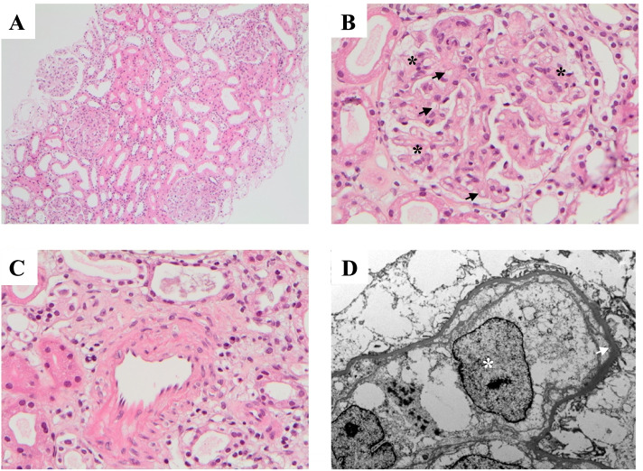
Fig. 2.
Renal Biopsy: A) Tubulointerstitium (H&E stain) - no significant interstitial fibrosis, tubular atrophy, or interstitial inflammation. B) Glomerulus (H&E stain) - mild expansion of mesangial matrix with patch endocapillary cellularity (black asterisks) and focal double contours (black arrows). No thrombi, sclerosing lesions, or necrotising lesions. C) Artery (H&E stain) – normal thickness with no fibrointimal hyperplasia or hylanosis. No evidence of arteritis, vasculitis or cholesterol emobli. D) Electron micrograph – swollen mesangial cells (white asterisks) with no electron-dense deposits or abnormal fibrils. Diffuse glomerular basement membrane thickening (white arrow). Tubules, interstitium, and vessels predominantly normal

