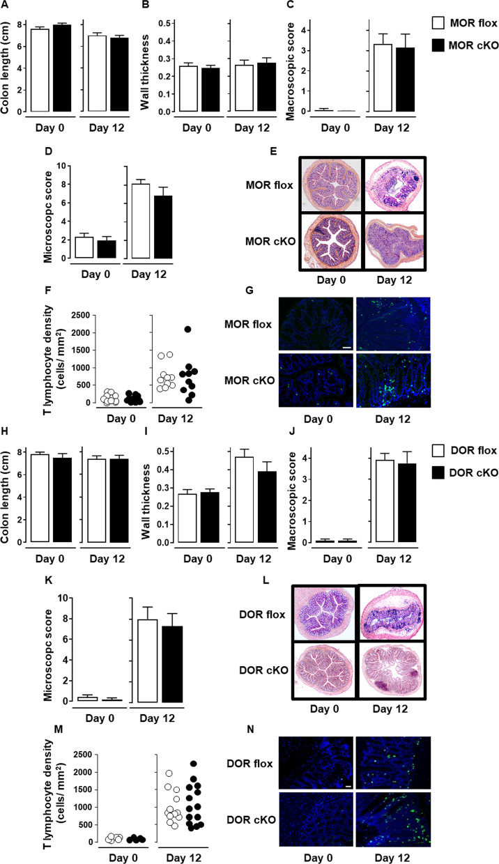
Fig. 4.
MOR or DOR deletion in Nav1.8-expressing sensory neurons does not alter neither colitis severity nor T cell infiltration within inflamed mucosa. Conditional knockout mice (black histogram) for either MOR (MOR cKO) (A–G) or DOR (DOR cKO) (H–N) were compared to their corresponding littermate wild-type floxed mice (MOR flox and DOR flox, respectively) (white histogram) in the basal conditions (Day 0) and on day 12 of DSS treatment for colitis severity (n = 7–8 mice/group) (A–E and H–L) and mucosal CD3+ T lymphocyte density (each point, corresponding to one animal, represents the mean of four different histological examinations per slice) (F, G; n = 10 mice/group and M, N; n = 6–14 mice/group). Representative histopathological H&E-stained colon sections (E and L) and anti-CD3 immunostaining (G and N) (scale 50 µm) at day 0 and day 12 of the DSS treatment for conditional MOR (E, G) and DOR (L, N) KO mice and their respective littermate wild-type MOR and DOR-floxed mice are shown. Data are expressed as mean ± SEM. Statistical analysis was performed using Mann–Whitney U test

