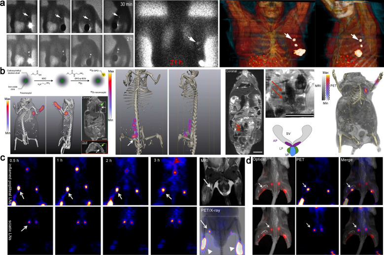
Fig. 3.
a The images obtained by intratumoral injection of 99mTc-Tilmanocept contrast agent in a patient with breast cancer. Two well-depicted SLNs were clearly seen in all sets of images (white arrow). Reproduced with permission from [85]. Copyright 2021, Society of Nuclear Medicine and Molecular Imaging. b Schematic of 89Zr-ferumoxytol and multimodal visualization of lymph nodes (axillary and somatic). Reproduced with permission from [87]. c The reconstructed coronal PET images of inflamed popliteal (Upper) and sciatic (Lower) LNs in the secondary hind limb inflammation model. d Complementary of optical (left) and PET (middle) images of enhancement popliteal and sciatic LNs, indicated by a white arrow. Reproduced with permission from [88]. Copyright 2014, Society of Nuclear Medicine and Molecular Imaging

