Fig. 4.
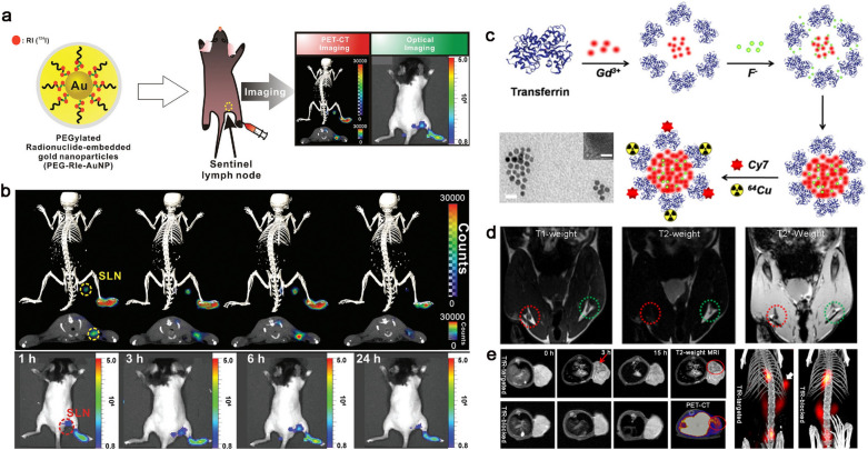
a Schematic illustration of PEG-RIe-AuNPs and multiple images of sentinel lymph nodes in vivo. b PET/CLI signal of PEG-RIe-AuNPs accumulate in sentinel lymph nodes over time. Detailed: (upper) 3D-PET/CT images, (middle) PET/CT images of cross-sectional, (bottom) CLI images. Reprinted with permission [89]. Copyright 2016, Wiley–VCH Verlag GmbH & Co. KGaA, Weinheim. c Self-assembly and modification of transferrin encapsulated GdF3 nanoparticles and TEM images. Scale bars are 100 nm (TEM) and 1 nm (HRTEM). d T1 and T2-weighted MRI images of SLNs in healthy mouse after injection of GdF3@Tf NPs 3 h. e Multimodal imaging of colorectal tumor bearing-mice at different time points after intravenous injection of 64Cu-GdF3@Tf-Cy7 NPs. Reproduced from [90]. Copyright 2020, American Chemical Society

