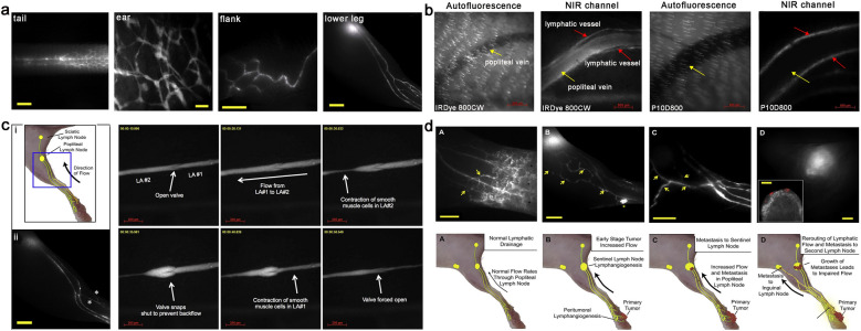
Fig. 8.
a Representative imaging of lateral side of lymphatic vessels in the tail, ear, flank and lower leg after intradermal injection of P20D680. b Specificity of tracer uptake by lymphatic vessels and popliteal vein after intradermal injection into hind paw with IRDye 800CW (ca.2 nm) and P10D800 (ca.7 nm). c The schematic diagram of lymphatic vessels network in mouse hind limbs (i) and the visualization of high uptake of P20D680 after intradermal injection into hind paw (ii). (iii) The valve function of collecting lymphatic vessels was also visualized in real time by P40D800. d Cartoon of lymphatic flow and rerouting of lymphatic flow after tumor spread from footpad tumors. Reproduced with permission from [124]. Copyright 2013, Elsevier

