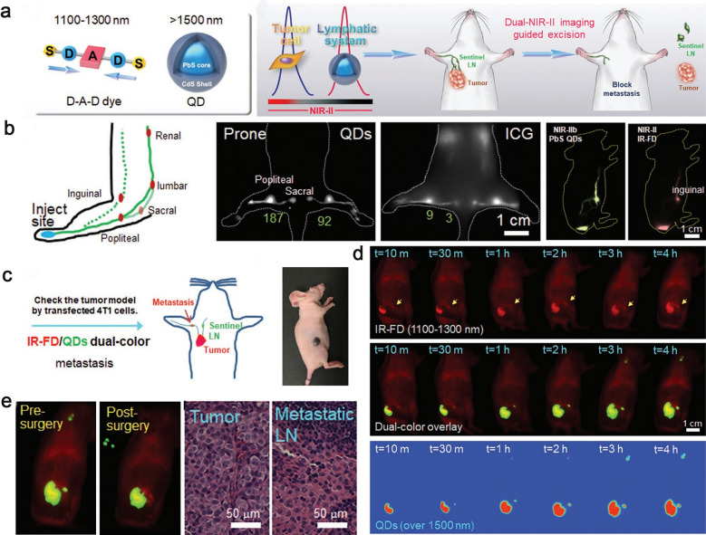
Fig. 9.
a General structure of dual NIR-II probes and NIR-II imaging-guided sentinel lymph nodes resection. b Schematic illustration of dual NIR II fluorescent dye injection and lymphatic drainage. c The illustration of dual-color imaging-guided surgery. d The different channels images displayed signal accumulation of IR-FD and QDs after administration of subcutaneous injection in tumor bearing mouse. e Dual NIR-II imaging guided pre- and post-surgery of sentinel LN excision. Reproduced with permission from [134]. Copyright 2020, Wiley–VCH Verlag GmbH & Co. KGaA, Weinheim

