Fig. 1.
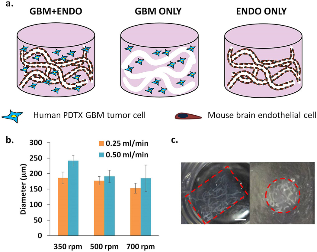
(a). Schematic of experimental group design. Endothelial cells (mouse brain) were patterned into vessellike structures in 3D biomimetic hydrogels using hydrolytically degradable alginate microfibers as porogens. In GBM + ENDO group, single patient-derived glioblastoma xenograft (PDTX GBM) cells were co-cultured with patterned endothelial cells. In GBM + ONLY group, single PDTX GBM cells were co-encapsulated with acellular alginate microfibers. In ENDO ONLY group, patterned endothelial cells were cultured alone. (b). Increasing calcium bath stir speed (350–700 rpm) led to decreased microchannel diameter. Increasing alginate injection rate (0.25, 0.50 mL/min) led to increased microchannel diameter. (c). Cross-sectional views of encapsulated alginate microfibers in 3D hydrogels. Left = side view. Right =top view. Red =hydrogel edge. Scale bar = 2 mm.

