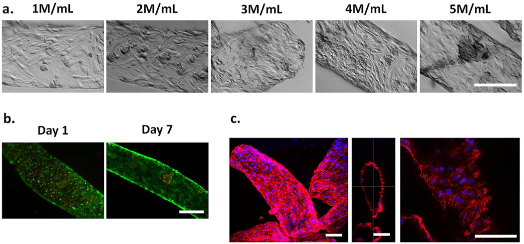
Fig. 2. Optimization of endothelial cell density in hydrolytically degradable alginate microfibers for microchannel formation in 3D hydrogels.
(a). Increasing initial endothelial cell density in alginate solution led to increased cell confluency and eventual cell aggregation after 2 days in culture. Scale bar = 250 μm. (b). Endothelial cells (4 M cells/mL) retained high cell viability and proliferated over time in microchannels. Scale bar =500 μm. Green =live. Red =dead. (c). Endothelial cells (4 M/mL) expressed high levels of CD31, along microchannel length and diameter after 7 days in culture. Scale bar = 100 μm. Blue =nuclei. Red = CD31. Maximal projection (left), orthogonal view (middle), single slice (right).

