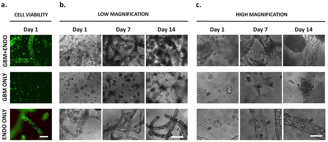
Fig. 3. Effects of co-culture on cell morphology over time.
(a). High cell viability of tumor and endothelial cells was observed after encapsulation. Scale bar =500 μm. Green = live. Red = dead. (b),(c). Endothelial cells started with rounded morphology after release from alginate microfibers on day 1. In GBM + ENDO group, endothelial cells were more rounded and disorganized, compared to cells in ENDO ONLY group, after 14 days in culture. Tumor cells started with rounded morphology after encapsulation. In GBM ONLY group, tumor cells formed large cell aggregates with radial protrusions after 14 days in culture. When co-cultured with endothelial cells in GBM + ENDO group, tumor cells directly adjacent to endothelial microchannels had more spherical morphology. Scale bars = 500 μm (b), 125 μm (c).

