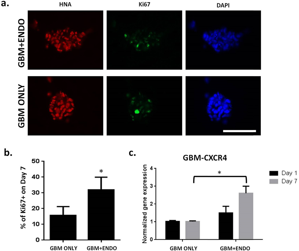
Fig. 5. Co-culture with endothelial cells led to increased tumor cell proliferation.
(a). Immunostaining for proliferating (Ki67+) tumor cells on day 7. Scale bar = 100 μm. Red = human nuclear antigen. Green = Ki67. Blue = nuclei. (b). Percentage of proliferating (Ki67+) tumor cells on day 7 (n = 150). * p < 0.05. (c). Expression of CXCR4 in tumor cells on day 7, normalized to Day 1. Human specific primers were used to measure gene expression levels of CXCR4 using RT-PCR. * p < 0.05. (For interpretation of the references to colour in this figure legend, the reader is referred to the Web version of this article.)

