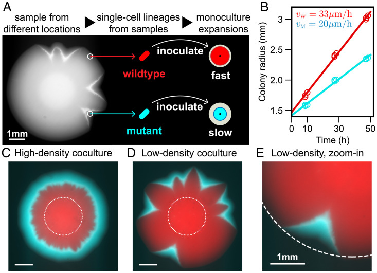
Fig. 1.
Slow mutant takes over the front with and without sector formation. (A) We found that wild-type R. planticola colonies develop V-shaped indentations; a bright-field image is shown. We sampled cells from the dents and nondented regions and then developed strains descending from a single cell (Materials and Methods). (B) The mutant expanded more slowly than the wild type when in isolation. The data points come from two technical replicates, and the line is a fit. (C and D) Despite its slower expansion on its own, the mutant wins in coculture. Fluorescence images show the spatial patterns 48 h after inoculation with a 99:1 mixture of the wild type and mutant. A ring of mutant (cyan) outrun and encircled wild type (red) when the mixed inoculant had a high density ( of ). Mutant sectors emerged and widened over the front when the mixed inoculant had a low density ( of ). Images are taken 48 h after inoculation, and dotted lines represent initial inoculant droplets. (E) A zoomed image of a V-shaped sector (from the bottom of D). Dotted circle is a fit from wild-type expansion. The advantage of the mutant and its slower expansion away from the wild type is evident from the lateral expansion of the cyan sector.

