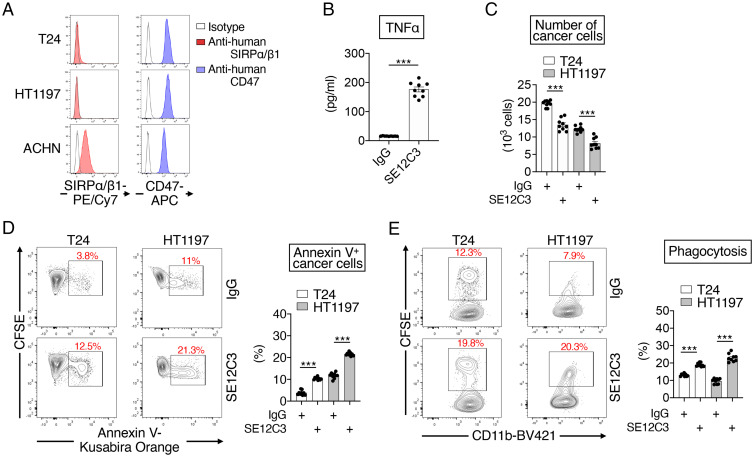
Fig. 7.
Promotion of macrophage-mediated killing of human bladder cancer cells by an mAb to human SIRPα/SIRPβ1. (A) Representative flow cytometric histograms for the expression of SIRPα or SIRPβ1 and CD47 on the cell surface of T24 and HT1197 human bladder cancer cells as well as on that of ACHN human renal cancer cells, which are positive for SIRPα expression (15). Data are representative of three separate experiments. (B) IFNγ-stimulated macrophages derived from human cord blood mononuclear cells were treated for 48 h with either control IgG or the SE12C3 mAb to human SIRPα/SIRPβ1 (each at 10 μg/mL), after which culture supernatants were harvested and assayed for TNFα. (C–E) CFSE-labeled T24 or HT1197 cells were cultured for 16 h with IFNγ-stimulated human macrophages in the presence of control IgG or SE12C3 (each at 10 μg/mL), after which the cells were collected for flow cytometric determination of the number of cancer cells (CFSE+CD11b–) (C), the proportion of annexin V+ cancer cells (annexin V+CFSE+CD11b–, apoptotic or dead) among all cancer cells (CFSE+CD11b–) (D), and the percentage of CFSE+CD11b+ macrophages (macrophages that had phagocytosed CFSE-labeled cancer cells) among all CD11b+ macrophages (E). Representative plots are shown (Left in D and E). All quantitative data are means ± SEM for three separate experiments, each performed in triplicate (n = 9 for each group) (B–E). ***P < 0.001 (two-tailed Welch’s t test).

