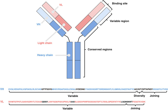
FIGURE 1.

Overview of the structure of an antibody and the sequence of its variable regions. An antibody contains two heavy (blue) and two light (red) chains, with each chain separable into one or more conserved (C) and one variable (V) region. The paired heavy and light V regions, annotated as VH and VL respectively, contain the binding site
