
FIGURE 2.
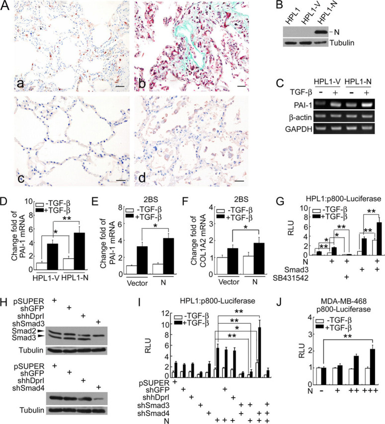
N protein enhances TGF-β-induced PAI-1 expression in a Smad3-dependent manner.A, immunohistochemistry staining shows the expression of N protein in SARS patient's lung (a), PAI-1 in normal lung (c), SARS patient's lung (d), and color developed by the 3-amino-9-ethylcarbazole and Masson's trichrome staining of collagen in SARS patient's lung (b); scale bar:50 μm. B–F, N protein up-regulates TGF-β target gene expression. The expression of N protein in stable HPL1 cells expressing N protein (HPL1-N) cells was determined by anti-N immunoblotting (B). The PAI-1 mRNA levels in HPL1-V or HPL1-N cells were analyzed by reverse transcription-PCR (C), or by real-time PCR (D). The PAI-1 mRNA levels (E) or COL1A2 (F) in 2BS cells transiently transfected with empty vector or N protein were analyzed by real-time PCR. β-Actin and GAPDH served as loading controls. G, N protein cooperates with Smad3 in enhancing the expression of luciferase driven by the PAI-1 promoter. HPL1 cells were co-transfected with p800-luciferase reporter (0.5 μg), pCS2-Myc-Smad3 (20 ng), and pcDNA3.1-N (0.5 μg) as indicated. At 24 h post-transfection, the cells were treated with 50 pm TGF-β with or without 10 μm SB431542. After 20 h, the cells were harvested for determination of luciferase activity. H, siRNAs efficiently knock down the expression of endogenous Smad3 and Smad4 in HPL1 cells. HPL1 cells were transfected with various siRNA constructs as indicated. After 0.2 mg/ml puromycin selection for 4 days, the cell lysates were collected and protein expression was determined by immunoblotting. pSR-shGFP and pSR-human Dapper I served as off-target siRNAs, and tubulin as the loading control. I, knockdown of Smad3 but not Smad4 in HPL1 cells represses the expression of TGF-β-induced p800-luciferase. HPL1 cells were co-transfected with p800-luciferase reporter, pSR-shGFP, pSR-human Dapper I, pSRG-shSmad3, or -shSmad4 and pcDNA3.1-N (0.5 μg for each construct) as indicated. Reporter assay was performed as in G. J, N protein enhances TGF-β-induced p800-luciferase expression in a dose-dependent manner in MDA-MB-468 cells. Cells were co-transfected with p800-luciferase reporter (0.5 μg) and pcDNA3.1-N (0.1, 0.3, or 0.5 μg). The asterisks indicate a statistically significant difference (*, p < 0.05; **, p < 0.01). RLU: relative luciferase units.