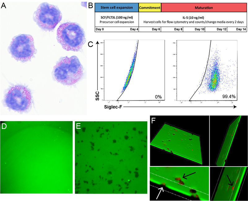
Figure 1. Bone marrow-derived murine eosinophils degrade fibrinogen substrates.
A) Day 14 IL-5-cultured eosinophils stained with a Diff-Quick Staining Kit and photographed with a 60x oil objective. Note mature red granular morphology. B) Schematic showing timeline of bone marrow-derived murine eosinophil culture protocol. C) Purity of cultured eosinophils, all cells were >99% Siglec-F positive (right) by non-stained control (left). D) FITC-linked fibrinogen substrate (green) without eosinophil addition visualized using fluorescent confocal microscopy. The substrate maintains a homogenous composition when eosinophils are not added, as is seen in this image. E) Dark spots represent areas of degraded fibrinogen after addition of eosinophils. F) Dark spots depict areas of total fibrinogen degradation, as visualized by using 3D stack images. Anti-MBP stained cells (red) localize to degraded areas and completely penetrate fibrinogen. Bottom two images are top images magnified, arrows point to anti-MBP stained cells penetrating the fibrinogen substrate. Note that cells tend to be smaller than the surrounding empty areas.

