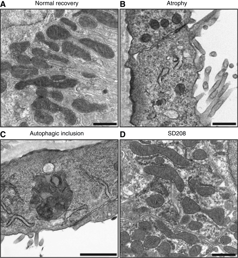
Figure 7.
Mitochondrial pathology in proximal tubules during the AKI to CKD transition. Electron micrographs showing (A) normal recovery, (B) atrophy with loss and simplification of mitochondria (C) autophagolysosome associated with atrophy and (D) TGF-β receptor antagonist, SD-208, improves mitochondrial number and morphology. Figure is from ref. (46) with permission.

