
Fig 5. Optical mapping of control and Cardiac-LKB1 KO mouse hearts.
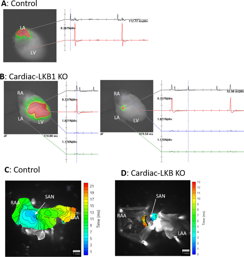
A: Representative image of a perfused control mouse heart. LA excitation was detected, and the heart was in sinus rhythm. B: Representative images of perfused Cardiac-LKB1 KO mouse hearts. No atrial excitation was detected and ventricular excitation was confirmed (left). The right image shows a focal area close to atrial septum that was excited independently and irregularly. C: Representative activation map of control mouse atria. Excitation was initiated from the SAN, then propagated though the RA and LA and ended in the RAA and LAA. D: A representative activation map of Cardiac-LKB1 KO mice atria. Excitation was observed only at SAN area; the excitation did not propagate through the atria.