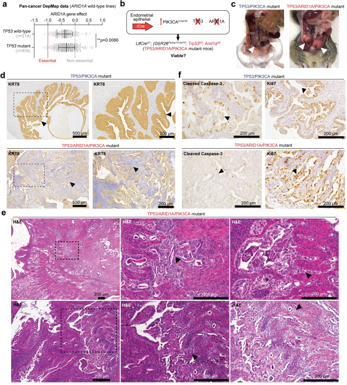
Fig 7. Co-existing TP53 and ARID1A mutations promote aggressive endometrial tumorigenesis.
a, Cancer dependency map (DepMap) data for ARID1A wild-type cell lines, measuring ARID1A knockout viability effect on TP53 wild-type vs. mutant lines. Statistic is unpaired, two-tailed Wilcoxon test. b, Schematic of genetically engineered mice harboring endometrial epithelial specific PIK3CAH1047R, TP53 loss, and ARID1A loss. c, Example gross necropsy images in TP53/PIK3CA mutant (top) and LtfCre0/+; (Gt)R26Pik3ca*H1047R; Trp53fl/fl; Arid1afl/fl (TP53/ARID1A/PIK3CA mutant, bottom) mice. Arrowheads denote uterine abnormalities. d, Immunohistochemical staining of KRT8 staining in TP53/PIK3CA and TP53/ARID1A/PIK3CA mutant uterus. Arrowheads denote mutant endometrial epithelial cells. In TP53/ARID1A/PIK3CA mutant image. e, Representative H&E uterine histology images of TP53/ARID1A/PIK3CA mutant mice. Arrowheads depict mutant tumor cells with squamous differentiation. f, Uterine IHC staining for Cleaved Caspase-3 (cell death, left) and Ki67 (proliferation, right) in TP53/PIK3CA mutant (top) and TP53/ARID1A/PIK3CA mutant (bottom) mice. Arrowheads denote endometrial epithelial cells.

