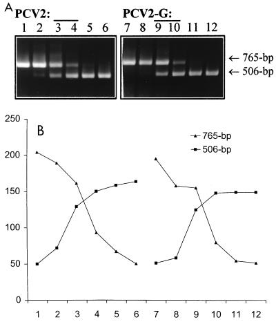
FIG. 2.
Application of cPCR for detection of PCV2 DNA in cell culture. (A) cPCR assay of PCV2 DNA in infected PCV-free PK-15 cells with or without d-glucosamine treatment. About 106 cells were infected with PCV2 at a multiplicity of infection of ∼1. One flask was incubated with 300 mM d-glucosamine for 30 min at 24 h postinfection. Both treated and untreated cells were harvested 24 h later. DNAs extracted from PCV2-infected cells (designated PCV2; lanes 1 to 6) and PCV2-infected, d-glucosamine-treated cells (designated PCV2-G; lanes 7 to 12) were coamplified with a selected range of 10-fold dilutions of competitor DNA (lanes 1 to 6 or 7 to 12, 1 ng, 100 pg, 10 pg, 100 fg, 10 fg, and 1 fg, respectively). (B) Quantification after densitometric scanning of the DNA bands in panel A. In both cases (PCV2 and PCV2-G) the equivalent point corresponds to 10 to 1 pg of competitor DNA.

