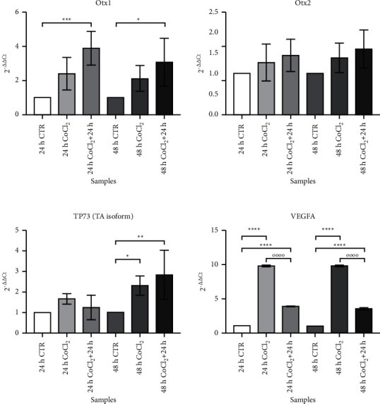
Figure 5.

qRT-PCR on CTR- and CoCl2-treated MIO-M1 Müller cells. Each graph represents the expression levels of a single studied gene in different experimental conditions. All values are indicated as median ± SD. On the Y-axis, the fold change value is reported. Statistical analysis was performed by one-way ANOVA and Tukey's post hoc test. ∗p < 0.05; ∗∗p < 0.01; ∗∗∗p < 0.001; and ∗∗∗∗p < 0.0001.
