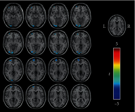
Figure 5.

Regions showing decreased FC in the tinnitus patients when the left precuneus was used as the seed (voxelwise P < 0.001, FWE-corrected clusterwise P < 0.05). In the tinnitus patients, the left inferior occipital gyrus, left calcarine cortex, and left superior frontal gyrus showed decreased FC with the left precuneus compared with those in the healthy controls. The color bar indicates the t value of independent two-sample t-tests between the groups. DAN: dorsal attention network; FC: functional connectivity; FWE: familywise error; L: left; R: right.
