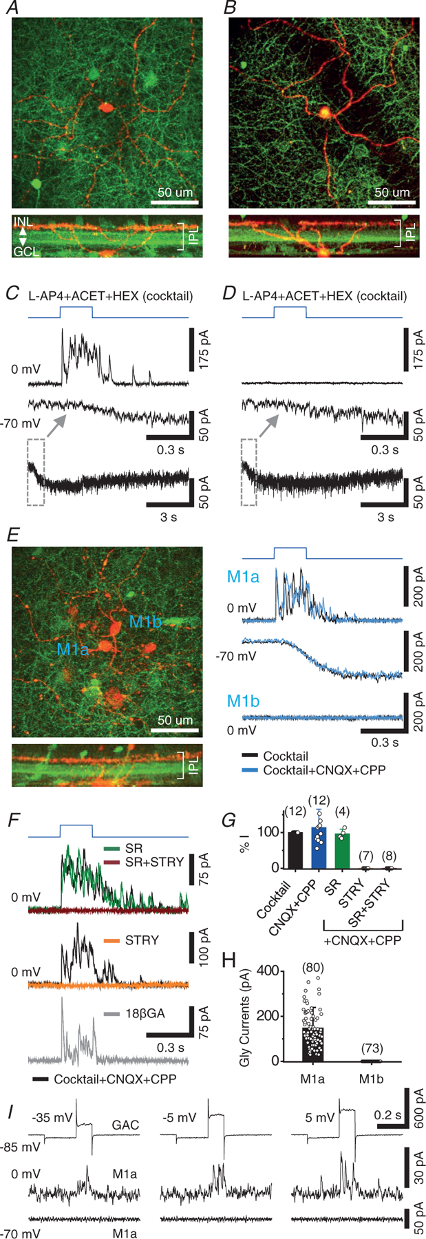
Figure 1. Selective glycinergic transmission from GACs to a subpopulation of M1 ipRGCs.

A, maximum z-projection (top) and cross-sectional reconstruction (bottom) of a two-photon image stack taken from a recorded M1 cell (red, Alexa 594) in a OPN4-GFP/vGluT3Cre/ChR2-YFP (green) retina. B, another example of a M1 cell. C, voltage clamp recording of the M1 cell in (A) in the presence of a control cocktail, showing fast outward postsynaptic currents at 0 mV (~Ecat) and slow melanopsin-mediated inward currents at −70 mV (~Ecl) in response to optogenetic activation of GACs by full-field blue light (blue trace). Bottom: slow inward currents on a longer time scale. D, voltage clamp recording of the M1 cell in (B), showing slow inward current at −70 mV, but no outward current at 0 mV. E, dual patch clamp recording of two adjacent M1 cells in the presence of control cocktail, showing robust outward postsynaptic currents in M1a cell (top), but no current in M1b cell (bottom), at 0 mV in response to optogenetic activation of GACs. The outward (top) and inward current (at −70 mV, middle) responses in M1a cell were resistant to CNQX (40 μm) + CPP (20 μm). F, top, middle: strychnine (STRY) (1–2 μm), but not gabazine (SR95531) (50 μm), completely abolished the outward responses of M1a cells at 0 mV in the cocktail + CNQX (40 μm) + CPP (20 μm). This optogenetically evoked glycinergic response in the cocktail remained largely intact after 15- to 25-min perfusion of 18 β-GA (bottom, 25 μm). G, summary of pharmacological effects on blue light (ChR2)-evoked peak outward currents (at 0 mV) in M1a cells. H, comparison of blue-light (ChR2)-evoked glycinergic current between M1a and M1b cells. I, dual patch clamp recording from a pair of GAC and M1a, showing voltage-gated currents of GAC (top) in response to depolarizing steps (100 ms long, preceded by 200 ms long pre-step from −85 mV to −100 mV) and outward postsynaptic current responses at 0 mV (middle), but no postsynaptic response at −70 mV (bottom), in the M1a cell. Values in parentheses indicate the number of cells tested. Error bars represent the SD. Cocktail concentrations: L-AP4 (20 μm), ACET (20 μm) and HEX (300 μm).
