
Figure 1:
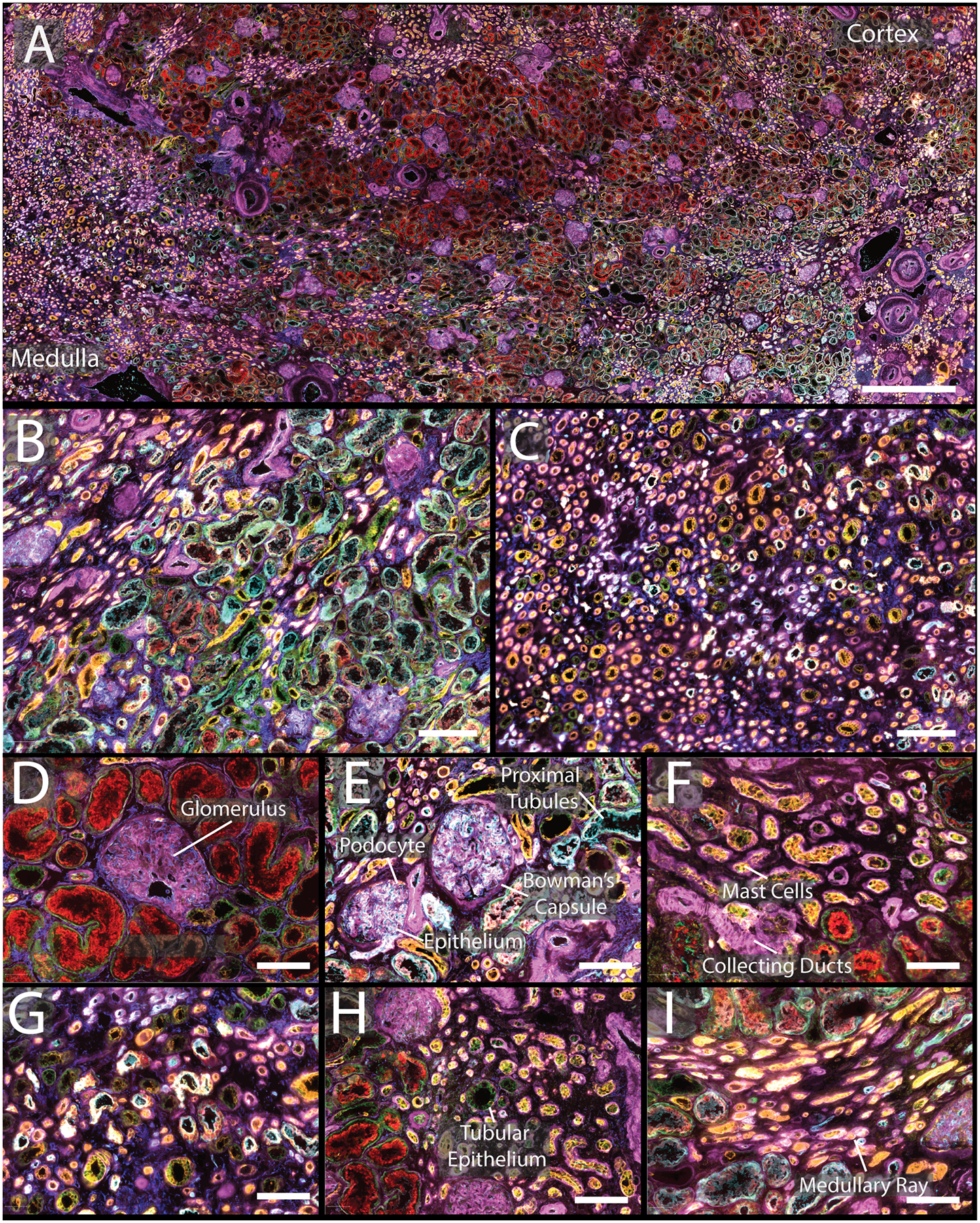
A) CODEX multiplexed IF staining of a human kidney from a 66-year-old male. B) Enlargement of a region of the cortex with several glomeruli present as well as proximal tubules. C) Medullary region of the kidney with many tubules present. D&E) Enlargement of glomeruli surrounded by different tubular segments with high expression of α-smooth muscle actin and aquaporin 1, respectively. F) Tubules within the cortex with high expression of cytokeratin 7 and tryptase. G) Representative view of medulla. H) Portion of cortex with increased numbers of mast cells. I) Enlargement of medullary rays. The color legend for all panels is as follows: cytokeratin 7 (pink), tryptase (orange), nestin (yellow), ß-catenin (green), aquaporin 1 (teal), vimentin (dark blue). Scale bars are 1 mm for panel A, 200 μm for panels B–C, and 50 μm for panels D–I.