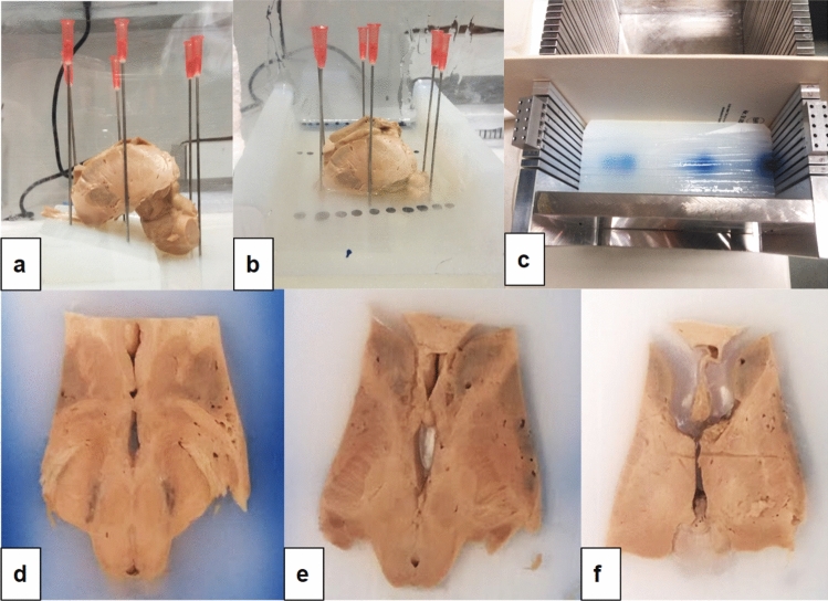
Fig. 17.
a, b, Pictures taken during the preparation procedure for cutting the midbrain specimen. The specimen is placed in a Perspex box (20 × 20 × 20 cm), with a titanium base and four mountable/ removable walls. b Approximately 2 L of a 5% agar solution was poured into the box, and when it started to set, the brain was placed on the agar base and the anterior commissure—posterior commissure was horizontally aligned; c the hardened agar block is placed in a box with rails of 5 mm spacing. d–f Examples of ACPC-parallel cuts of 5 mm thickness (macro, unstained)

