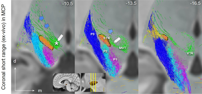
Fig. 6.
Short range ex-vivo depiction of PFC fibers connecting to STN (via CP, P1–P3, lateral) and ventral tegmentum (via lh, medial), coronal view. Close proximity of P1–P3 with STN. P1 fibers traverse the nucleus. White arrow marks the region in MVT/PRF identical with “limbic STN cone” in Haynes and Haber (2013). Blue arrows point to motorMFB fibers in Zi. Insets show the position of coronal cuts. Zi zona incerta; MVT mesencephalic ventral tegmentum; VTA ventral tegmental area; * VTA terminal field (approximated))

