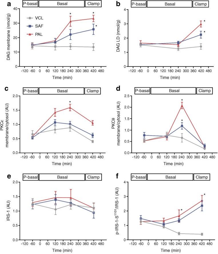
Fig. 5.
Myocellular lipid metabolites and insulin signalling (DAG–nPKC pathway) in healthy humans. DAG species 18–1:18–1, 16–0:16–0 and 16–0:18–1 in the cell membrane fraction (a), DAG species 18–1:18–1, 16–0:16–0 and 16–0:18–1 in the lipid droplet fraction (b), nPKCε activation (c), nPKCθ activation (d) and IRS-1 levels (e) as well as serine1101-phosphorylation of IRS-1 relative to IRS-1 (f) during the pre-basal, basal and clamp periods after ingestion of PAL (red), SAF (blue) or VCL (water, grey) at 0 min. Expression signals on immunoblots are expressed in arbitrary units (AU) after normalising against GAPDH for total and cytosolic proteins and against Na+/K+-ATPase for membrane proteins. Data are shown as means ± SEM; n = 16 at time point −60 min, n = 10 at +120 min, n = 6 at +240 min and +420 min. *p < 0.05 vs VCL at same time point (ANOVA adjusted for repeated measures with Tukey–Kramer correction for each time point between interventions). LD, lipid droplet fraction; P-basal, pre-basal

