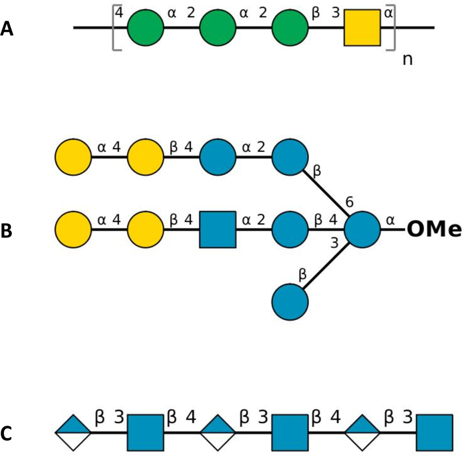
Figure 2.

Symbol representation of carbohydrate sequences of (A) E. coli O176 O-antigen repeating unit (n = 10), (B) M. catarrhalis serotype C oligosaccharide, and (C) hyaluronan hexasaccharide in PDB ID 1LOH: blue circle for d-glucose (Glc), green circle for d-mannose (Man), yellow circle for d-galactose (Gal), blue square for N-acetyl-d-glucosamine (GlcNAc), yellow square for N-acetyl-d-galactosamine (GalNAc), and half-filled blue diamond for d-glucuronic acid (GlcA).
