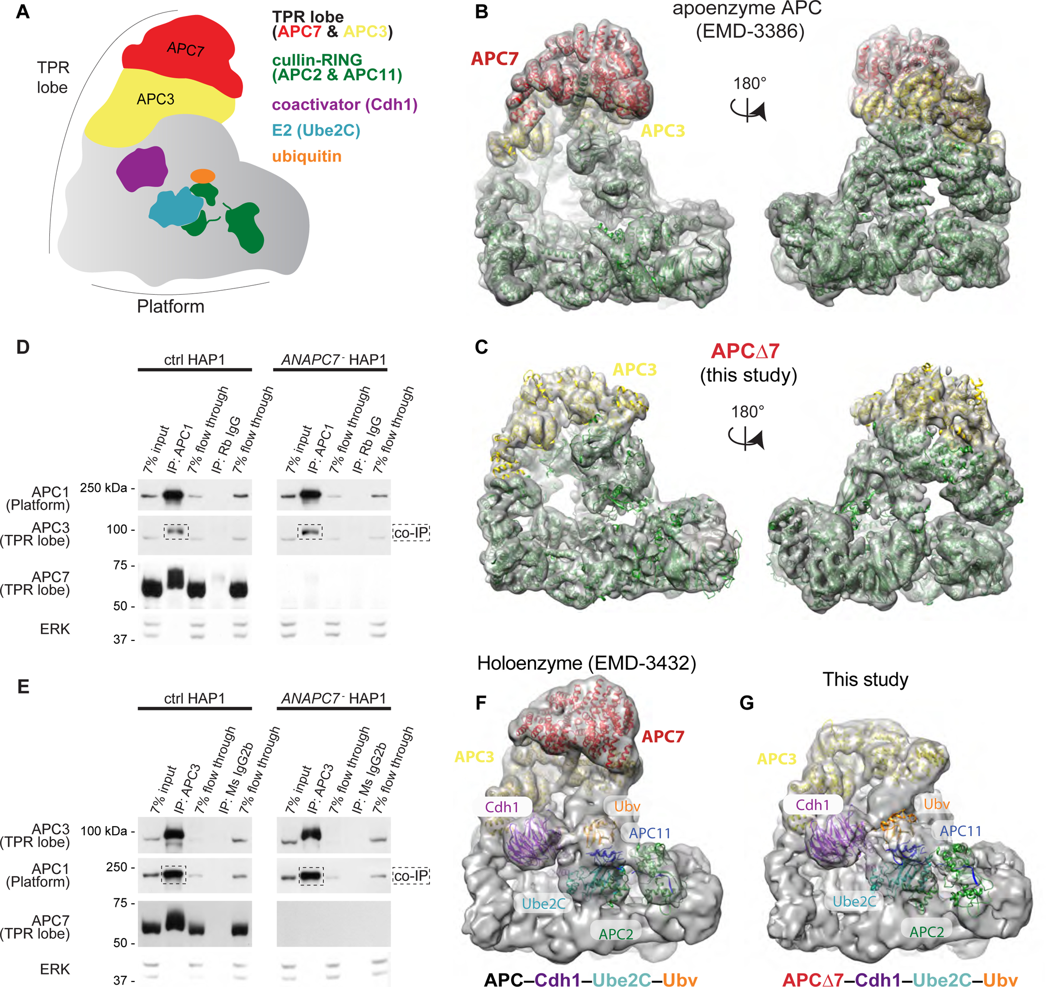
Figure 1: APC7 is not required for the conformation of the APC or its assembly. See also Figure S1.

A. Schematic of the APC.
B. Published model of wild-type human APC (EMD-3386) showing the APC7 dimer (Chang et al., 2015).
C. Empirically determined Cryo-EM structure of APCΔ7. APC subunits from published coordinates (PDB 4L9U) were rigid-body docked using Chimera (Pettersen et al., 2004).
D. IP of APC1 from HAP1 cells followed by immunoblot (IB) for APC subunits. Flow through fractions reflect the remaining protein after antibody binding.
E. IP of APC3 (TPR lobe) from HAP1 cells followed by IB for APC subunits.
F. Published Cryo-EM structure of the APC holoenzyme (APC–Cdh1–Ube2C–substrate–Ubv) trapped by chemical crosslinking (Brown et al., 2016).
G. Cryo-EM structure of APCΔ7–Cdh1–Ube2C–substrate–Ubv with Ube2C poised for ubiquitin transfer. Published coordinates (PDB 4L9U) were rigid-body docked using Chimera.
