
Figure 6: APC7 operates in the context of Cdh1-APC to drive Ki-67 degradation in neurons.
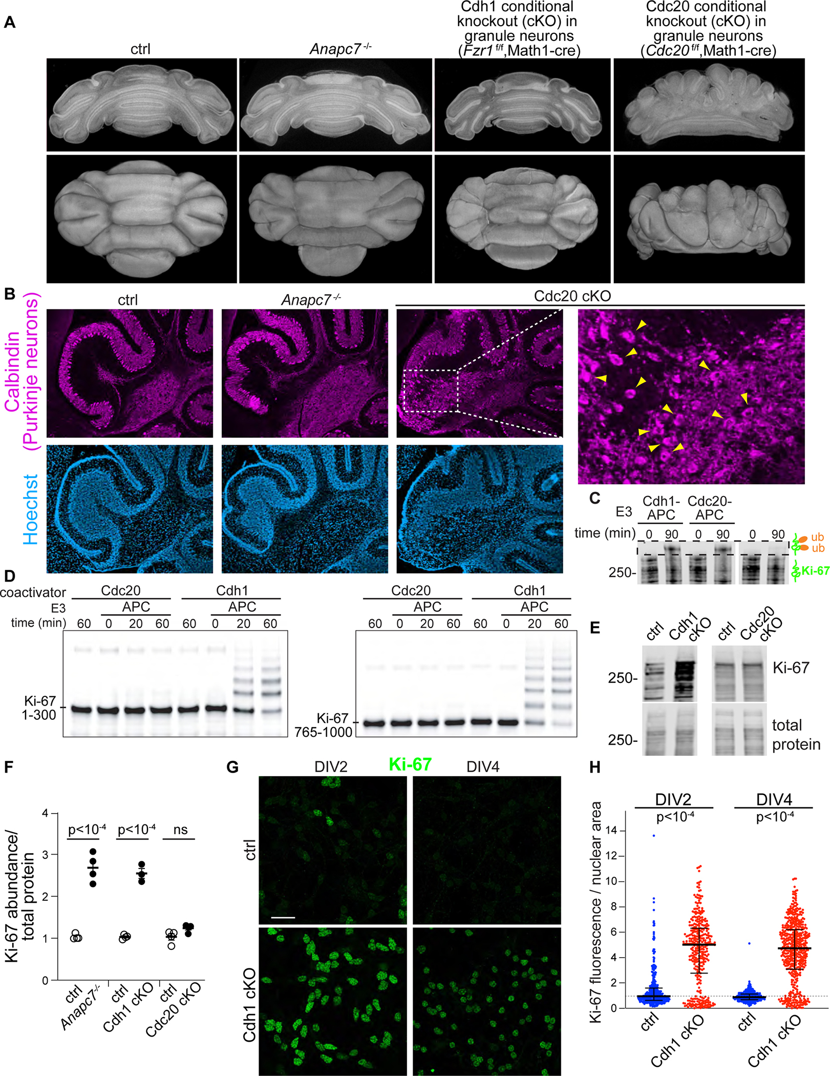
A. Electron tomography of P8 cerebellum.
B. Calbindin IF in sagittal sections of P12 cerebellum. An expanded region from the Cdc20f/f,Math1-cre is shown (PCL, Purkinje cell layer).
C. In vitro ubiquitination of full-length Ki-67 by recombinant Cdh1-APC and Cdc20-APC. Ki-67 was immunoprecipitated from wild-type P7 cerebellum and detected by Ki-67 IB.
D. In vitro ubiquitination of human Ki-67 amino acids 1–300 (left) and 765–1000 (right) by Cdh1-APC and Cdc20-APC.
E. Ki-67 IB in P12 cerebellum.
F. Densitometry quantitation of Ki-67 IB. Mutants were normalized and compared to littermates. Error bars SEM (N = 4, p-value by two-tailed unpaired t-test).
G. IF of Ki-67 in primary cerebellar granule neuron cultures.
H. Quantitation of Ki-67 IF normalized to nuclear area in cultured neurons on the indicated DIV. Error bars interquartile range (p-values by Kruskal-Wallis test for non-parametric data followed by Dunn test).