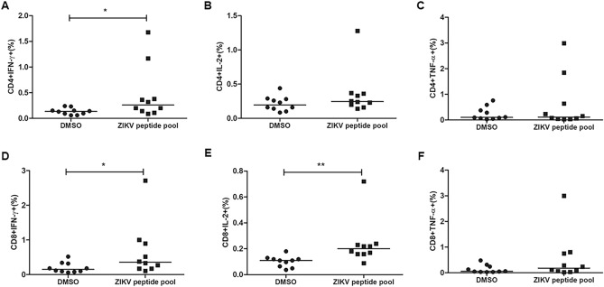
Figure 7.
CD4 + and CD8 + T cell responses to ZIKV peptides. PBMC of 10 individuals with previous infection to ZIKV were stimulated for 5 h with the ZIKV peptide pool. After 1 h of stimulation, Brefeldin A was added. The cells were stained with antibodies for the surface markers CD3, CD4 and CD8 and intracellular stained with anti-IFN-γ, IL-2 and anti-TNF-α. IFN-γ, IL-2 and TNF-α production by CD4 + T cells (A–C) and CD8 + T cells (D–F) was evaluated by flow cytometry. The horizontal lines indicate the medians. Mann–Whitney test was performed to determine statistical significance and p < 0.05 was considered significant.

