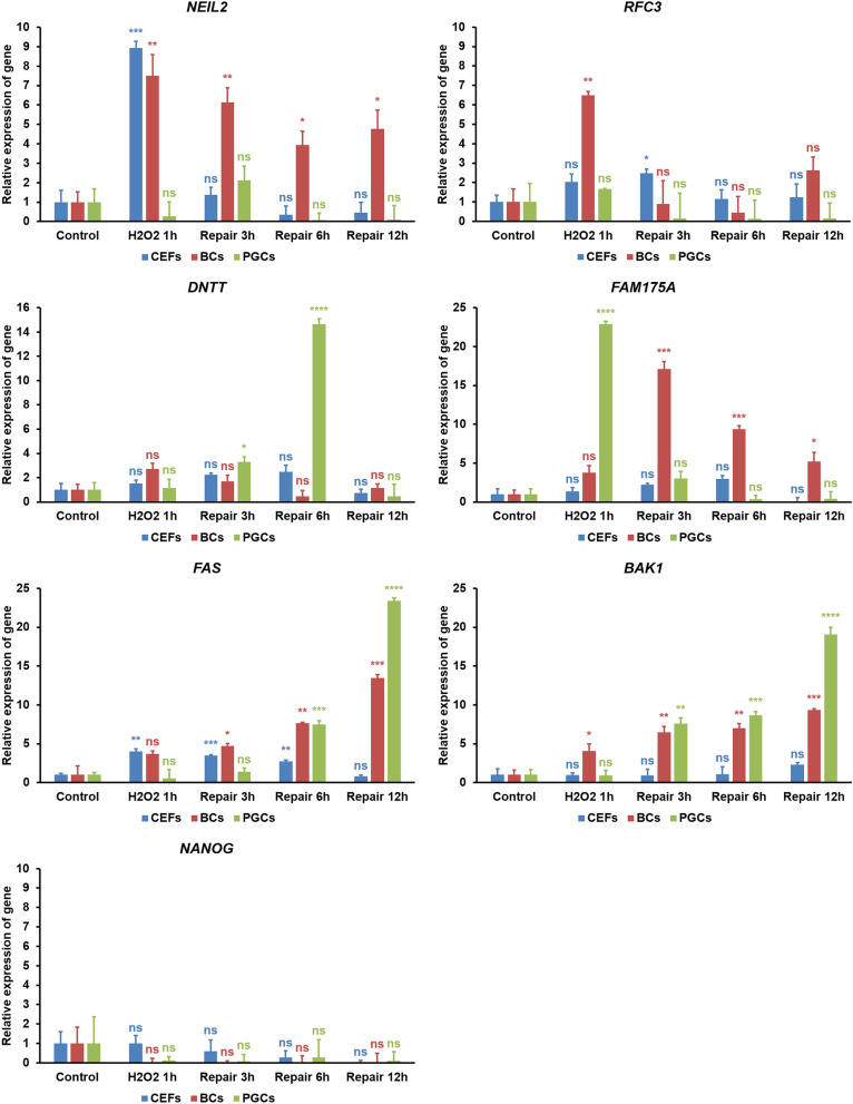
Figure 8.
RT-qPCR analysis in CEFs, EGK.X blastoderm cells (BCs), and PGCs treated with H2O2. The cultured CEFs, EGK.X blastoderm cells, and PGCs were treated with 1 mM H2O2 for 1 h (H2O2 1 h) in one group. In three other groups, the cells were treated with 1 mM H2O2 for 1 h, and then incubated in fresh media without H2O2 for 3 h (repair 3 h), 6 h (repair 6 h) and 12 h (repair 12 h). After treatment, cDNA of the cells was prepared and amplified with specific qPCR primers of the candidate genes from the BER pathway (NEIL2), NER/MMR pathway (RFC3), NHEJ pathway (DNTT), HR pathway (FAM175A), apoptosis pathway (FAS and BAK1), and pluripotency regulating pathway (NANOG). The relative expression of genes was normalized with the chicken GAPDH and respective control sample, and analyzed by the 2−ΔΔCt method. Significant differences between the respective control and treated samples were determined by Student’s t test. Statistical significance was ranked as *P < 0.05, **P < 0.01, ***P < 0.001, or ****P < 0.0001. ns, non-significant.

