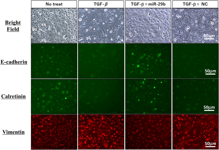
Figure 2.
Phenotypic changes of human peritoneal mesothelial cells (HPMC) by stimulation with TGF-β1. HPMC were cultured with 10 ng/ml TGF-β1 for 48 h. In some wells, HPMC were transfected with miR-29b-3p mimic or negative control (NC) with lipofectamine RNAiMAX at a final concentration of 50 nM, and cultured with TGF-β1 for 48 h at 37 °C. Morphological changes by light microscopy and expression of E-cadherin, calretinin and vimentin were observed with a fluorescein microscopy, BZ-X710 (Keyense, Osaka, JAPAN). Magnification: ×400. All the figures were generated using BZ-H3A software (Keyense, Osaka, JAPAN) (https://www.keyence.com/products/microscope/fluorescence-microscope/bz-x700/models/bz-h3ae/).

