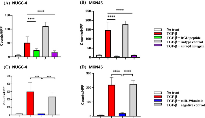
Figure 4.
Adhesion of gastric cancer cells on human peritoneal mesothelial cells (HPMC). HPMC were cultured with TGF-β as described in the legend of Fig. 2 and then fluorescein labelled NUGC-4 and MKN45 were added, incubated for 15 min on the mesothelial cell (MC) monolayer. After gentle washing with warmed media for 3 times, the number of NUGC-4 cells remaining attached were counted under a fluorescent microscope. In (A) and (B), MC monolayer was incubated with RGD peptide (40 μM), anti-β1 integrin mAb or isotype control mouse IgG (20 μg/ml) before the addition of tumor cells. In (C) and (D), MC monolayer was pretreated with miR-29b mimic or negative control as described in the legend of Fig. 2. Data show mean ± standard deviation in 1 of the 2 different experiments ***p < 0.001, ****p < 0.0001.

