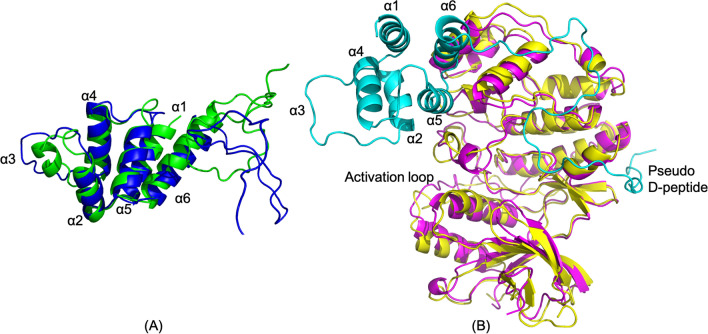
Figure 2.
The simulated complex structure of PEA-15/ERK2. (A) PEA-15 in the complex (green) is superimposed with its free form (blue) with the RMSD 2.734 Å for the DED. Helix α3 is uncoiled and shifts in α2 and α4 can also be observed. (B) ERK2 in the complex is superimposed with the crystal structure of 4IZ5 with the RMSD 2.002 Å. The overall backbone of ERK2 does not show any significant difference between the simulated and crystal structure. The molecular structures were visualized and generated using PyMOL version 2.4 (http://www.pymol.org/).

