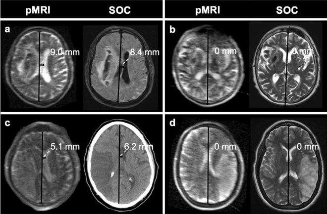
Figure 2.
Example midline shift measurements on portable MRI (pMRI) and standard-of-care (SOC) imaging exams. (a) 81-year-old male with right intracerebral hemorrhage. Midline shift was measured to be 9.0 mm and 8.4 mm on the pMRI T2-weighted (T2W) and standard-of-care (SOC) MRI fluid-attenuated inversion recovery (FLAIR) images, respectively. (b) 43-year-old male with right intracerebral hemorrhage. No midline shift was measured on either pMRI T2W or SOC MRI T2W exams. (c) 71-year-old male with right M1 occlusion. Midline shift was measured to be 5.1 mm and 6.2 mm on the pMRI T2W and SOC CT images, respectively. (d) 44-year-old female with left M2 occlusion. No midline shift was measured on either pMRI T2W or SOC MRI T2W exams. Figure created using: Microsoft PowerPoint, Version 16.52, https://www.microsoft.com/en-us/microsoft-365/powerpoint.

