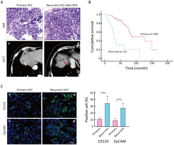
Figure 1.
Enrichment of TICs in recurrent tissues after IRFA treatment. (A) Representative H&E staining (a,b) and CECT images of primary HCC (white arrow in c) and recurrent HCC (red arrow in d), * in d indicating the necrotic lesion of RFA treatment; (B) Overall survival rate for primary and recurrent HCC patients after RFA was compared using Kaplan-Meier analysis (P<0.05); (C) Immunofluorescence staining was used for detecting the expression of CD133+ and EpCAM+ TICs in primary and recurrent HCC tumors. Samples were obtained in seven patients with both primary tumors and recurrent tumors. Positive cells were stained with green signal; nuclei were stained with DAPI (blue). The bar graph in the right indicates the proportion of Ki-67+ and CD34+ cells as
 . TIC, tumor-initiating cell; IRFA, insufficient radiofrequency ablation; H&E, hematoxylin & eosin; CECT, contrast-enhanced computed tomography; HCC, hepatocellular carcinoma; RFA, radiofrequency ablation; EpCAM, epithelial cell adhesion molecule; DAPI, 4’,6-diamidino-2-phenylindole.***, P<0.001.

